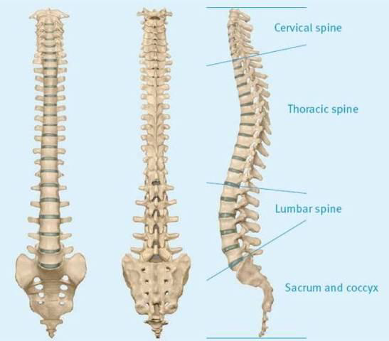
Parts of vertebral column is a bony structure that extends from the foramen magnum (the hole at the bottom of the skull where the brain is joined to the spinal cord) to the sacral hiatus. The vertebral column consists of 7 cervical, 12 thoracic, and 5 lumbar vertebrae as well as the sacrum, and coccyx (Figure 6:1). The first cervical vertebra is called the atlas. The atlas has a unique anatomical structure that allows for articulations to the base of the skull and second cervical vertebrae. The second cervical vertebra is called the axis. Each of the 12 thoracic vertebrae articulates with a corresponding rib.
The bony vertebral column provides:
On a lateral view the vertebral canal exhibits four curvatures (Figure 6:1), of which the thoracic convexity (kyphosis) and the lumbar concavity (lordosis) are of major importance to the distribution of local anesthetic solution in the subarachnoid space.
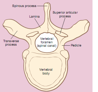
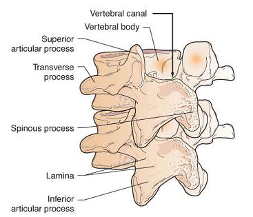
Although the vertebrae show regional differences they all conform to a basic pattern: the body and the vertebral (or neural) arch which surrounds and protects the spinal cord lying in the vertebral foremen.
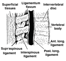
Ligaments maintain the shape of the vertebral column and provide support (Figure 6:4). Vertebral bodies and disks are connected and supported on the ventral side by anterior and posterior longitudinal ligaments. On the dorsal side of the vertebral column the ligamentum flavum, interspinous ligament, and supraspinous ligament provide support. These dorsal ligaments are structures that the anesthesia provider will pass through when placing a needle for neuraxial blockade. With experience the anesthesia provider will be able to identify these structures through tactile feel.